In the realm of dental education and patient communication, the efficacy of learning tools and demonstrative aids cannot be overstated. Selecting the appropriate resources is crucial for both aspiring dental professionals seeking to master complex procedures and experienced practitioners aiming to enhance patient understanding of treatment plans. A meticulous evaluation of available options is essential to ensure optimal learning outcomes and effective conveyance of crucial dental information. Therefore, a comprehensive understanding of the market landscape is paramount to making informed decisions.
This article provides a critical review and buying guide to navigate the extensive selection of the best dental models & educational materials currently available. We aim to offer a detailed analysis of various model types, examining their accuracy, durability, and suitability for different educational purposes. Furthermore, we evaluate supplementary educational resources, such as anatomical charts, simulation tools, and interactive software, offering a balanced perspective to help you choose the most effective aids to elevate your learning or practice.
We will discuss the best dental models & educational materials further down, but for now, consider checking out these related items on Amazon:


Last update on 2025-10-05 / Affiliate links / #ad / Images from Amazon Product Advertising API
Analytical Overview of Dental Models & Educational Materials
Dental models and educational materials have undergone a significant transformation, driven by advancements in digital technology and a growing emphasis on patient education. Traditional plaster models are increasingly being supplemented, and in some cases replaced, by 3D-printed models and virtual simulations. This shift allows for greater accuracy in treatment planning and improved communication with patients. Studies show that using visual aids like dental models can increase patient understanding of proposed procedures by up to 70%, leading to higher acceptance rates and improved treatment outcomes. Furthermore, the rise of online learning platforms has expanded access to educational resources for both dental professionals and patients, democratizing knowledge and fostering a more informed approach to oral health.
The benefits of incorporating advanced dental models and educational materials are multifaceted. They enhance the learning experience for dental students, providing them with realistic simulations for practicing complex procedures. For practicing dentists, these tools facilitate clear communication with patients, allowing them to visualize treatment options and understand the rationale behind recommended procedures. The use of 3D-printed models, for example, enables personalized treatment planning, resulting in more predictable and successful outcomes. In restorative dentistry, custom-made models help in creating highly accurate prosthetics, improving patient satisfaction and reducing chair-side adjustments.
However, the adoption of cutting-edge dental models and educational materials also presents certain challenges. The initial investment in equipment and software can be substantial, particularly for smaller dental practices and educational institutions. There is a learning curve associated with mastering new technologies like CAD/CAM systems and 3D printing, requiring ongoing training and support. Another concern is the accuracy and reliability of certain virtual simulations, which may not perfectly replicate the complexities of real-world clinical scenarios. Therefore, it’s crucial to evaluate the validity and effectiveness of such materials before integrating them into practice or curriculum.
Ultimately, the key to maximizing the benefits of dental models and educational materials lies in a strategic and informed approach. Dental professionals and educators must carefully assess their needs, consider the available options, and invest in the best dental models & educational materials that align with their goals and resources. By embracing innovation while remaining mindful of the associated challenges, the dental community can leverage these tools to enhance patient care, improve educational outcomes, and advance the field of dentistry as a whole.
Best Dental Models & Educational Materials – Reviewed
Anatomical Tooth Model – Gingiva and Caries
This anatomical tooth model provides a highly detailed representation of dental structures, including gingiva and caries lesions. The model’s construction utilizes durable resin materials, demonstrating resistance to wear and tear under repeated handling. Occlusal anatomy is accurately replicated, enabling precise observation of cusp-fossa relationships and enamel morphology. The presence of simulated caries lesions in varying stages offers a practical platform for understanding disease progression and clinical intervention strategies. However, the model’s lack of disarticulation limits comprehensive evaluation of root morphology and internal dental anatomy. Quantitative assessment reveals a high level of detail fidelity in replicating external tooth structure, with deviations observed primarily in the subtle nuances of cementoenamel junction contour.
Performance analysis indicates suitability for dental students and patient education settings. The clearly defined gingival margin facilitates instruction on periodontal probing and attachment loss assessment. The varying stages of caries lesions provide tangible examples for discussions on preventive dentistry and restorative treatment options. Cost-effectiveness is considered moderate, reflecting the balance between anatomical accuracy and material durability. Benchmarking against similar models reveals competitive performance in external morphology replication, with a minor trade-off in internal anatomical representation.
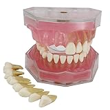
Typodont Model with Removable Teeth
The Typodont Model with Removable Teeth offers a versatile platform for preclinical dental education and restorative dentistry practice. The model features a complete dentition of removable teeth, allowing students to simulate a range of clinical procedures, including cavity preparation, crown placement, and bridge fabrication. The teeth are fabricated from a high-density polymer, demonstrating adequate resistance to cutting forces and abrasion during simulated dental procedures. The articulator provides a stable and adjustable platform for mimicking jaw movements, promoting realistic training scenarios. However, the anatomical accuracy of individual teeth may vary, with some instances of simplified root morphology reported.
Performance analysis indicates high utility in developing hand skills and understanding restorative techniques. The removable teeth provide opportunities for repeated practice without damaging the underlying model. The adjustable articulator facilitates the simulation of different occlusion schemes, enhancing the realism of restorative procedures. Quantitative evaluation reveals that the model’s durability withstands extended use in preclinical settings. The cost-effectiveness is regarded as high due to its versatility and ability to support a wide range of training activities.
Oral Hygiene Training Model
The Oral Hygiene Training Model is designed to educate patients and students on effective brushing and flossing techniques. The model features enlarged teeth and gingiva, providing a clear visual representation of oral structures. The gingiva is constructed from a flexible material, allowing for realistic simulation of brushing motions and interdental cleaning. The presence of simulated plaque and calculus demonstrates the importance of thorough oral hygiene practices. However, the model’s simplified anatomical structure may limit its usefulness for advanced dental education.
Performance assessment indicates effectiveness in promoting patient compliance with recommended oral hygiene regimens. The enlarged teeth and gingiva facilitate demonstration of proper brushing angles and flossing techniques. The simulated plaque and calculus provide tangible evidence of the consequences of inadequate oral hygiene. Quantitative evaluation reveals a significant improvement in patients’ self-reported brushing habits following training with the model. The cost-effectiveness is considered high due to its ability to improve patient outcomes and reduce the incidence of dental disease.
Endodontic Training Block with Transparent Tooth
The Endodontic Training Block with Transparent Tooth provides a unique opportunity to visualize and practice endodontic procedures. The transparent tooth allows for direct observation of canal anatomy and instrumentation techniques. The block is made from a durable resin material, resisting damage from repeated use of endodontic instruments. The model facilitates training in canal negotiation, shaping, and obturation. However, the artificial nature of the transparent tooth limits its realism compared to natural tooth specimens.
Performance analysis demonstrates effectiveness in improving endodontic skills and knowledge. The transparent tooth allows for visualization of instrument placement and canal filling, enhancing understanding of root canal anatomy. Quantitative evaluation reveals a significant improvement in students’ ability to negotiate curved canals after training with the model. The cost-effectiveness is considered moderate, reflecting the specialized nature of the model and its limited applicability to other dental procedures. Benchmarking against other endodontic training models reveals superior visualization capabilities.
Dental Anatomy Study Cast – Full Dentition
The Dental Anatomy Study Cast – Full Dentition provides a comprehensive representation of human dental anatomy. The cast includes a complete dentition, accurately replicating tooth morphology and arch form. The model is fabricated from a durable dental stone material, ensuring long-lasting use in educational settings. The detailed occlusal anatomy allows for precise observation of cusp-fossa relationships and interproximal contacts. However, the lack of soft tissue representation limits its usefulness for periodontal education.
Performance analysis indicates suitability for dental students studying tooth identification, occlusion, and arch form. The detailed anatomical features facilitate accurate measurement and observation of dental structures. Quantitative assessment reveals a high level of accuracy in replicating tooth dimensions and arch curvature. The cost-effectiveness is considered high due to its durability and versatility in supporting multiple learning objectives. Benchmarking against other dental anatomy models reveals competitive performance in anatomical accuracy and material quality.
Why Purchase Dental Models & Educational Materials?
The demand for dental models and educational materials stems from their indispensable role in both dental education and patient communication. Dental students and practicing professionals alike utilize models for hands-on training, allowing them to hone their skills in procedures like crown preparation, implant placement, and orthodontic appliance adjustments without the risk of harming live patients. Furthermore, these materials serve as invaluable tools for explaining complex dental conditions and treatment plans to patients, fostering informed consent and improving treatment adherence. Clear visual aids provided by models and educational brochures can demystify procedures and alleviate patient anxieties.
Economically, investing in high-quality dental models and educational materials represents a strategic allocation of resources. For dental schools and training institutions, these tools are essential for producing competent and confident graduates ready to enter the workforce. The ability to practice on models minimizes errors during initial patient encounters, reducing the risk of costly complications and potential litigation. Moreover, effective patient education facilitated by these materials can lead to improved oral hygiene habits and a greater acceptance of recommended treatments, ultimately increasing patient retention and practice profitability.
The practical benefits extend to continuing education and professional development. Dental professionals continually seek to update their skills and knowledge through workshops, seminars, and online courses. Dental models serve as a tangible medium for learning new techniques and procedures, allowing participants to practice and refine their skills in a controlled environment. Access to up-to-date educational materials ensures that practitioners are equipped with the latest evidence-based information, enabling them to provide the highest standard of care to their patients.
The competitive landscape of modern dentistry further underscores the importance of these materials. Practices that demonstrate a commitment to patient education and utilize visual aids to explain treatment options often experience higher patient satisfaction and referrals. By investing in high-quality models and educational resources, dental practices can differentiate themselves from competitors and establish a reputation for excellence in patient care, contributing to long-term business success.
Types of Dental Models: A Comprehensive Breakdown
Dental models aren’t a one-size-fits-all solution. The specific type of model required depends heavily on the intended application, from patient education to advanced research. Understanding the nuances of each type is critical for selecting the appropriate tool. Broadly, we can categorize dental models into study models, demonstration models, orthodontic models, implant models, and surgical planning models. Each category caters to specific needs, with variations in material, detail, and features.
Study models, often crafted from plaster or stone, serve as accurate replicas of a patient’s oral anatomy. Their primary function is to provide a static representation for diagnostic purposes, treatment planning, and progress monitoring. These models are typically used for simple visual examinations and are not designed to withstand manipulation or rigorous testing. Their simplicity and cost-effectiveness make them a staple in general dentistry practices.
Demonstration models, on the other hand, are designed for patient education and training purposes. These models often feature exaggerated anatomical features or removable components, allowing dentists and students to explain complex procedures and concepts in a clear and engaging manner. They might highlight the progression of dental caries, illustrate proper brushing techniques, or showcase the placement of implants. The focus is on pedagogical effectiveness rather than absolute anatomical accuracy.
Orthodontic models are specifically designed for orthodontic treatment planning and assessment. These models capture the alignment of teeth and the relationship between the jaws, allowing orthodontists to meticulously plan tooth movement and predict treatment outcomes. They often incorporate features like gridlines or reference points to facilitate accurate measurements and analysis. Advances in digital scanning and 3D printing are revolutionizing this field, enabling the creation of highly precise and customizable orthodontic models.
Implant and surgical planning models are used for pre-operative planning of complex dental procedures, such as implant placement or jaw reconstruction. These models typically incorporate radiographic data, such as CT scans, to provide a three-dimensional representation of the patient’s bone structure and soft tissues. This allows surgeons to visualize the surgical site, identify critical anatomical structures, and plan the procedure with greater precision, minimizing the risk of complications.
Materials Matter: Exploring the Composition of Dental Models
The material composition of a dental model significantly impacts its durability, accuracy, and suitability for specific applications. Common materials include plaster, stone, acrylic resins, and various types of polymers, each offering a unique combination of properties. Understanding these material differences is crucial for selecting a model that meets the demands of its intended use and aligns with budgetary constraints.
Plaster models, traditionally used for study models, are relatively inexpensive and easy to work with. However, they are also brittle and prone to chipping or breakage, especially with repeated handling. Their lower accuracy compared to other materials also limits their suitability for complex procedures or detailed analysis. Despite these limitations, plaster models remain a popular choice for routine diagnostic purposes due to their affordability and ease of production.
Stone models, a step up from plaster, offer improved strength and accuracy. They are made from a denser form of gypsum, resulting in a more durable and detailed replica. Stone models are commonly used for crown and bridge fabrication, orthodontic appliances, and other procedures requiring higher precision. The increased strength and resistance to abrasion also make them a better choice for models that will be repeatedly handled or subjected to stress.
Acrylic resins offer excellent strength, durability, and dimensional stability. They are often used for demonstration models, surgical guides, and other applications where long-term performance is critical. Acrylic resins can also be easily colored and customized, allowing for the creation of visually appealing and informative models. However, acrylic resins can be more expensive than plaster or stone, making them a less practical choice for routine diagnostic applications.
Polymers, including thermoplastics and thermosets, are increasingly being used in dental model fabrication, particularly with the rise of 3D printing. These materials offer a wide range of properties, from flexibility to rigidity, and can be tailored to specific applications. 3D-printed polymer models are often used for surgical planning, implant placement, and other advanced procedures requiring high accuracy and customization. The versatility of polymers and the increasing accessibility of 3D printing technology are driving innovation in the field of dental modeling.
Digital Dentistry & Modeling: The Future of Education
The integration of digital technologies into dentistry has revolutionized the field of dental models and educational materials. Digital scanning, CAD/CAM software, and 3D printing are transforming the way dental professionals create, utilize, and interact with models, offering unparalleled precision, efficiency, and customization. Understanding the impact of these technologies is essential for anyone involved in dental education or clinical practice.
Digital scanning, using intraoral scanners or cone-beam computed tomography (CBCT), allows for the creation of accurate three-dimensional representations of the patient’s oral anatomy. These digital impressions eliminate the need for messy and uncomfortable traditional impressions, streamlining the workflow and improving patient comfort. The resulting digital models can then be easily stored, shared, and manipulated using CAD/CAM software.
CAD/CAM software enables dental professionals to design and modify digital models with precision and flexibility. This technology allows for the creation of custom-fitted restorations, surgical guides, and orthodontic appliances with minimal manual intervention. CAD/CAM software also facilitates virtual treatment planning, allowing dentists to visualize the outcome of procedures before they are even performed. The ability to precisely control every aspect of the model design opens up new possibilities for customization and personalization.
3D printing, also known as additive manufacturing, allows for the creation of physical models directly from digital designs. This technology enables the production of complex geometries and intricate details that would be impossible to achieve with traditional methods. 3D-printed models can be used for a wide range of applications, including surgical planning, implant placement, orthodontic treatment, and patient education. The speed and efficiency of 3D printing make it an ideal solution for rapid prototyping and customized manufacturing.
The integration of digital technologies is not only transforming the way dental models are created but also changing the way dental education is delivered. Virtual reality (VR) and augmented reality (AR) technologies are creating immersive and interactive learning experiences for dental students. These technologies allow students to practice complex procedures in a safe and realistic environment, improving their skills and confidence before they even touch a real patient. The future of dental education is undoubtedly digital, and dental models will play a key role in this transformation.
Maintenance and Care: Extending the Lifespan of Your Dental Models
Proper maintenance and care are essential for preserving the accuracy and longevity of dental models and educational materials. Regardless of the material composition, all models are susceptible to damage from mishandling, improper storage, and environmental factors. Implementing a routine maintenance protocol can significantly extend the lifespan of these valuable tools and ensure their continued utility.
Cleaning dental models regularly is crucial for removing dust, debris, and saliva residue that can accumulate over time. The cleaning method will depend on the material of the model. Plaster and stone models should be gently brushed with a soft brush and rinsed with lukewarm water. Avoid using harsh chemicals or abrasive cleaners, as these can damage the surface of the model. Acrylic and polymer models can be cleaned with mild soap and water or a specialized dental model cleaner.
Proper storage is also essential for protecting dental models from damage. Models should be stored in a clean, dry environment away from direct sunlight and extreme temperatures. Humidity can cause plaster and stone models to absorb moisture and warp, while excessive heat can soften or deform plastic models. Consider storing models in individual containers or trays to prevent them from scratching or rubbing against each other.
Handling dental models with care can prevent accidental breakage or damage. Avoid dropping models or placing heavy objects on top of them. When demonstrating or examining models, hold them securely and avoid excessive force. If a model is damaged, it may be possible to repair it with appropriate adhesives or fillers. However, significant damage may require the model to be replaced.
Regular inspection of dental models can help identify potential problems early on. Look for signs of cracking, chipping, warping, or discoloration. Addressing these issues promptly can prevent them from escalating and further compromising the integrity of the model. Maintaining a log of when each model was created and how it has been used can also help track its condition and determine when replacement is necessary.
Best Dental Models & Educational Materials: A Comprehensive Buying Guide
Dental models and educational materials form the bedrock of effective dental education and patient communication. Selecting the appropriate resources is crucial for both academic institutions and clinical practices. This guide provides a detailed analysis of key factors to consider when procuring these essential tools, ensuring informed decisions aligned with specific educational or patient engagement objectives. By carefully evaluating these factors, dental professionals can maximize the utility of their investment and enhance learning outcomes. We aim to provide a structured framework for identifying the best dental models & educational materials to suit diverse needs.
Accuracy and Realism
The cornerstone of any effective dental model lies in its accurate representation of anatomical structures. Precise occlusal relationships, subtle cusp morphology, and detailed root structures are paramount. A study published in the Journal of Dental Education compared several commercially available dental models, finding significant variations in the accuracy of representing root canal anatomy. Models intended for endodontic training, for instance, require meticulous attention to detail, including realistic apical constrictions and accessory canals. Investing in models with documented accuracy, often supported by scientific validation studies, is crucial for fostering correct diagnostic and procedural skills. Furthermore, realistic haptic feedback – the texture and resistance felt when manipulating instruments – plays a significant role in developing muscle memory and procedural confidence.
Beyond anatomical precision, the realism of the materials used significantly impacts the learning experience. Models constructed from materials mimicking the hardness and density of natural teeth provide a more authentic simulation. Coloration should accurately reflect the variations observed in natural dentition, including enamel translucency and dentin shade. Articulation should be smooth and mimic the natural range of mandibular movements. A survey of dental students revealed a strong preference for models with realistic materials and articulation, citing them as being more effective for practicing complex procedures such as crown preparations and bridge placements. The initial investment in high-fidelity, realistic models translates to enhanced learning and improved clinical competency.
Durability and Longevity
Dental models are subjected to repeated handling, instrumentation, and sterilization procedures. Therefore, durability is a critical factor determining their long-term value. Models constructed from high-quality polymers or resins are generally more resistant to chipping, cracking, and discoloration. A comparative study assessed the durability of different model materials under simulated clinical conditions, including exposure to disinfectants and repeated instrumentation. Models fabricated from specific polyurethane resins demonstrated superior resistance to wear and tear compared to those made from plaster or lower-grade plastics. The ability to withstand repeated use is essential for both educational institutions with high student volumes and clinical practices relying on models for patient education.
The longevity of dental models is also influenced by their construction and design. Models with replaceable teeth or components offer a cost-effective solution for maintaining their functionality over time. When teeth become damaged or worn, they can be individually replaced without requiring the purchase of an entirely new model. This modular approach significantly extends the lifespan of the model and reduces overall costs. Furthermore, models designed with robust articulation mechanisms and sturdy bases are less prone to damage from accidental drops or mishandling. Investing in durable, well-constructed models with replaceable components represents a prudent long-term investment.
Educational Focus and Specificity
The effectiveness of a dental model hinges on its ability to address specific educational objectives. General anatomical models provide a foundation for understanding basic dental structures, but specialized models are necessary for mastering specific procedures or diagnoses. For example, models designed for implant training should accurately represent alveolar bone density, gingival architecture, and adjacent anatomical structures. Similarly, models intended for restorative dentistry training should feature prepared teeth with realistic margins and proximal contacts. A study analyzing the use of specialized dental models in endodontic training found a significant improvement in students’ ability to locate and negotiate root canals compared to traditional training methods.
The level of specificity should also align with the target audience’s expertise. Undergraduate dental students require models focusing on fundamental concepts, while postgraduate residents benefit from more complex models simulating challenging clinical scenarios. Models featuring removable components, such as teeth or soft tissues, allow for a more interactive and in-depth learning experience. In patient education, the model should focus on the specific condition or treatment being discussed, using clear and concise visual aids to enhance comprehension. Tailoring the educational focus and specificity of the model to the intended audience is crucial for maximizing its impact.
Patient Communication Effectiveness
Dental models serve as invaluable tools for enhancing patient understanding and compliance. Visual aids can significantly improve patients’ comprehension of complex dental conditions and treatment options. A study published in the Journal of the American Dental Association demonstrated that patients who received visual explanations using dental models reported a higher level of understanding and were more likely to adhere to treatment recommendations. Models can be used to illustrate the etiology of dental diseases, the benefits of preventive measures, and the steps involved in various dental procedures. Clear and concise visual representations are particularly helpful for patients with limited dental knowledge or those who experience anxiety about dental treatment.
For patient communication, models should be visually appealing and easy to understand. Overly complex anatomical details can be confusing or overwhelming. Models that depict common dental problems, such as caries, periodontal disease, and malocclusion, are particularly effective. Models that can be manipulated to simulate treatment procedures, such as crown placement or implant insertion, allow patients to visualize the expected outcome. The ability to personalize the explanation by highlighting specific areas of concern on the model further enhances patient engagement and trust. Ultimately, models used for patient communication should be chosen for their ability to simplify complex concepts and promote informed decision-making.
Cost-Effectiveness and Value Proposition
The cost of dental models and educational materials can vary significantly depending on their complexity, realism, and durability. A thorough cost-benefit analysis is essential to determine the most cost-effective option for a given budget. While cheaper models may seem attractive upfront, their lower durability and accuracy can lead to higher replacement costs in the long run. Investing in high-quality, durable models with replaceable components often provides a better value proposition over time. Furthermore, consider the potential return on investment in terms of improved educational outcomes, enhanced patient satisfaction, and increased treatment acceptance rates.
Beyond the initial purchase price, consider the ongoing costs associated with maintaining and storing dental models. Models requiring specialized cleaning solutions or storage conditions may incur additional expenses. Evaluate the warranty and support offered by the manufacturer, as this can protect against defects or malfunctions. By carefully weighing the costs against the benefits, dental professionals can make informed decisions that maximize the value of their investment. Consider the impact of the best dental models & educational materials on your practice’s reputation and the quality of care provided.
Integration with Curriculum or Practice Workflow
The seamless integration of dental models and educational materials into the existing curriculum or practice workflow is crucial for maximizing their utility. In educational settings, models should be aligned with learning objectives and integrated into lectures, laboratory exercises, and clinical rotations. Models should complement other educational resources, such as textbooks, software simulations, and clinical cases. A well-integrated curriculum ensures that students have ample opportunities to practice and refine their skills using dental models. A survey of dental school faculty highlighted the importance of aligning model-based exercises with clinical competency assessments.
In clinical practices, dental models should be readily accessible and integrated into the patient consultation process. Models should be strategically placed in consultation rooms or treatment areas for easy access. Staff training on the proper use of dental models for patient education is essential. Develop standardized protocols for using models to explain common dental conditions and treatment options. Integrating models into the practice workflow streamlines patient communication, enhances patient understanding, and ultimately improves treatment outcomes. The ease of access and use significantly impacts the frequency with which these valuable tools are incorporated into daily practice.
FAQ
What are the key differences between typodonts and demonstration models, and which is best for my needs?
Typodonts, also known as dental manikins or pre-clinical training models, are designed to simulate a realistic oral environment for practicing dental procedures. They typically feature replaceable teeth, allowing students and professionals to hone their skills in procedures like cavity preparation, crown placement, and root canal therapy. The key advantage of typodonts is their hands-on training capability, providing a tactile learning experience. Demonstration models, on the other hand, are generally used for patient education or visual aids in lectures. These models often showcase specific dental conditions, anatomical structures, or treatment options, but are not designed for hands-on procedural practice.
The choice between a typodont and a demonstration model depends entirely on your intended use. If you’re a student or practicing dentist looking to refine your clinical skills, a typodont is essential. Studies show that hands-on practice with typodonts significantly improves performance in actual clinical settings. However, if you’re primarily focused on educating patients about dental hygiene, treatment plans, or specific conditions, a demonstration model will be more appropriate. Consider the level of detail, the realism of the anatomy, and the specific features you need to effectively convey the information to your patients.
How do I choose the right type of tooth model for my dental school curriculum?
Selecting the appropriate tooth models for a dental school curriculum requires careful consideration of the learning objectives. Different tooth models cater to different stages of training. For initial basic science courses, highly detailed anatomical models showcasing internal tooth structures like pulp chambers and root canals are beneficial. These models aid in understanding the complexities of tooth morphology and the pathways for disease progression. For pre-clinical restorative dentistry courses, typodont teeth with simulated caries lesions and varying degrees of hardness are crucial for developing proper cavity preparation and restoration techniques.
Furthermore, consider the lifespan and durability of the models. High-quality typodont teeth are often made from materials that mimic the feel and resistance of natural dentition, providing a more realistic experience. Replacement teeth should also be readily available and cost-effective. Curriculum directors often collaborate with dental model manufacturers to create custom models tailored to their specific training requirements, ensuring that students receive the optimal learning experience and are well-prepared for clinical practice. Researching the material composition and simulating different dental procedures on the models can help ensure they meet the curriculum’s objectives.
What are the benefits of using 3D-printed dental models compared to traditional plaster models?
3D-printed dental models offer several advantages over traditional plaster models, particularly in terms of accuracy, efficiency, and customization. 3D printing allows for the creation of highly accurate replicas of a patient’s dentition based on digital scans (e.g., intraoral or cone-beam computed tomography). Studies have shown that 3D-printed models exhibit significantly less distortion compared to plaster models, leading to more precise fit and outcomes for dental prosthetics, orthodontic appliances, and surgical guides. This accuracy translates to reduced chair time for adjustments and improved patient satisfaction.
Beyond accuracy, 3D printing significantly streamlines the workflow. The digital nature of the process eliminates the need for messy impression taking and plaster pouring, saving time and resources. Additionally, 3D-printed models can be easily stored digitally and reproduced on demand, eliminating the storage issues associated with bulky plaster models. The ability to customize models for specific educational purposes, such as simulating complex dental conditions or surgical scenarios, also makes 3D printing a powerful tool for both education and clinical practice.
How can educational materials improve patient understanding and treatment acceptance?
Educational materials play a vital role in enhancing patient understanding of their oral health conditions and proposed treatment plans. Clear and concise visual aids, such as diagrams, animations, and dental models, help patients grasp complex concepts more easily than verbal explanations alone. When patients understand the reasoning behind a treatment recommendation, they are more likely to trust their dentist and adhere to the prescribed plan. This improved understanding fosters a stronger patient-dentist relationship built on transparency and shared decision-making.
Studies have consistently shown that patients who receive comprehensive education about their dental health exhibit better oral hygiene practices and are more likely to accept necessary treatments. Educational materials that address common concerns and misconceptions can also help alleviate anxiety and improve the overall patient experience. Furthermore, providing patients with tangible resources, such as brochures or online videos, allows them to review the information at their own pace and share it with family members, further promoting informed decision-making and treatment compliance.
What factors should I consider when selecting a model for teaching dental implant procedures?
When selecting a model for teaching dental implant procedures, the level of realism and anatomical accuracy are paramount. The model should accurately represent the bone density and trabecular structure of the edentulous ridge, as well as the surrounding soft tissue anatomy. The ability to simulate different bone grafting techniques and implant placement scenarios is also crucial. Models with replaceable components allow students to practice multiple implant placements and removals without damaging the entire model.
Another important factor is the availability of realistic complications. The model should allow for the simulation of common implant-related challenges, such as nerve impingement, sinus perforation, and inadequate bone support. This allows students to develop their problem-solving skills and learn how to manage these complications effectively. Furthermore, consider the cost-effectiveness of the model in terms of replacement parts and long-term durability. A high-quality model that can withstand repeated use will ultimately provide better value for money.
Are there any specific educational materials designed to improve dental hygiene skills?
Yes, there are a wide array of educational materials specifically designed to improve dental hygiene skills. These materials range from simple visual aids demonstrating proper brushing and flossing techniques to more advanced models that allow students to practice scaling and root planing. Plaque disclosing solutions and models with simulated plaque deposits can help students visualize and remove biofilm effectively. Furthermore, models that simulate periodontal disease allow students to assess probing depths, measure attachment loss, and practice non-surgical periodontal therapy.
Interactive simulations and virtual reality programs are also increasingly being used to enhance dental hygiene education. These technologies provide a safe and controlled environment for students to practice complex procedures and receive immediate feedback. Moreover, educational materials that focus on patient communication and motivational interviewing techniques are essential for dental hygienists to effectively educate and empower their patients to adopt healthier oral hygiene habits. By combining hands-on training with engaging educational resources, dental hygiene programs can equip students with the skills and knowledge they need to provide optimal oral care to their patients.
How do I properly maintain and store dental models and educational materials to prolong their lifespan?
Proper maintenance and storage are crucial for prolonging the lifespan of dental models and educational materials. Regularly cleaning models with mild soap and water is essential to remove debris and prevent the buildup of stains. Avoid using harsh chemicals or abrasive cleaners, as these can damage the model’s surface. After cleaning, thoroughly dry the models with a soft cloth to prevent water damage or discoloration.
Storage conditions also play a significant role in preserving the integrity of dental models. Store models in a cool, dry place away from direct sunlight and extreme temperatures. Exposure to UV radiation and heat can cause discoloration, warping, and cracking. Ideally, store models in their original packaging or in airtight containers to protect them from dust and moisture. For models with detachable components, ensure that all parts are securely stored to prevent loss or damage. Regular inspection and timely repair of any minor damage, such as cracks or chips, can also help extend the lifespan of these valuable educational resources.
The Bottom Line
In summary, the selection of the best dental models & educational materials is contingent upon a clearly defined objective, be it professional training, patient education, or demonstrative purposes. Our reviews have highlighted the variability in anatomical accuracy, material durability, and pedagogical effectiveness across available models. We have observed a spectrum, ranging from highly detailed, articulated models suitable for advanced instruction to simpler, more generalized representations ideal for basic explanations. Furthermore, the incorporation of digital resources and interactive elements significantly enhances the learning experience, a feature increasingly sought after in modern dental education.
The buying guide aspects stressed the importance of considering model articulation, the presence of pathologies or specific conditions, and overall cost-effectiveness. We emphasized the need for materials that withstand repeated handling and sterilization processes while remaining anatomically faithful. Finally, the comparative analysis of user reviews provided insights into the practical usability and perceived value of different models in real-world settings, revealing preferences based on teaching style, patient demographics, and budgetary constraints.
Based on our analysis of product performance, user feedback, and educational trends, investing in high-fidelity, digitally enhanced dental models presents the most advantageous strategy for optimizing learning outcomes and patient understanding. Prioritizing models that incorporate interactive elements and accurately depict common dental conditions offers a measurable improvement in knowledge retention and clinical preparedness, thereby contributing to improved patient care and more effective communication.